Fear and the amygdala

What is fear? Why are we sometimes afraid? Can fear be inhibited? What produces fear – the brain or the heart?

It is definitely the brain! More exactly, something in the brain – a tiny, almond-shaped structure, which sits anteriorly to the hippocampus, called the amygdala. This small part of our brain is to blame for  the perception of fearful stimuli and the physiological responses (increased heart rate, electrodermal responses etc.) to fearful stimuli.

As part of the FEAR system, the amygdala connects to the medial hypothalamus and the dorsal periaqueductal grey matter (in the midbrain), which is important in pain modulation in the dorsal horn of the spinal cord, as well as to sensory and association cortices. The lateral nucleus of the amygdala receives inputs from different brain regions, thus allowing the formation of associations, required for aversive conditioning. Following the processing in the lateral nucleus, information about the stimulus is, then, projected to the central nucleus of the amygdala, where an appropriate response to the stimuli is initiated, provided that the stimuli are detected as threatening or potentially dangerous.

The amygdala is involved in emotional learning and memory, modulating implicit learning, explicit memory, attention, social responses, emotion inhibition and vigilance.

You can find the article on memory here, to brush up a bit.

  • Implicit memory is a type of learning, which cannot be voluntarily reported or remembered. It includes the memories for skills and habits, for procedural knowledge, grammar and languages, priming, simple forms of associative learning and classical conditioning. The latter is particularly important for fear. It involved a conditioned stimulus (CS) and an unconditioned, painful/fearful stimulus (US), with US preceding CS and determining a fearful response to CS. This type of fear learning is adaptive and is known as sensitisation or acquisition.

There are two different pathways in the amygdala, important for fear conditioning. The “low road” pathway: sensory information projects to the thalamus, which directly communicates with the amygdala; this pathway is fast, modulating rapid responses of the amygdala to different types of fearful stimuli. The “high road” pathway is an indirect pathway: sensory information projects to the thalamus and from there, it is conveyed to the sensory cortex for a finer analysis; the sensory cortex, then, communicates the processed information to the amygdala. This pathway ensures that the sensory stimulus is the conditioned stimulus. So the responses of amygdala to threatening stimuli are both rapid and sure.

  • Explicit memory refers to the memory of facts and events; in the case of fear is means the processing and retention for a long time, of emotional events and information. For this type of fear learning, the amygdala interacts with the hippocampus. There is a distinction between the formation of memory for aversive experience (fear conditioning), which is based on previous experience, and explicit learning (in the hippocampus), which involves learning and remembering aversive properties of different threatening stimuli. The memory in the hippocampus is enhanced by arousal produced in the amygdala. The activation of the amygdala can make different cortical areas, not just the hippocampus, more receptive to stimuli that are adaptively important, thus ensuring that unattended, but important stimuli gain access to consciousness.
  • Social responses involve the ability to recognise a stimulus as good, bad, neutral or arousing. This ability, however, does not depend on the amygdala. There is one exception here, otherwise we wouldn’t be talking about it in the context of fear mediated by amygdala – fearful facial expressions. According to Darwin, social species, like humans, use facial expressions to detect internal emotional states of other members of the group. This function, mediated by the amygdala, is crucial in the emotional regulation of human social behaviour. Damage to amygdala has been demonstrated to result in impairment of the patient to identify fearful faces correctly and, therefore, react to them, accordingly. It should also be noted there is no need for the subject’s awareness of the fearful stimulus, for the amygdala to respond. In other words, when a fearful facial expression is presented subliminally, the amygdala will still show activation.
  • Inhibition of fear – it is actually very difficult to escape your own fears, as fear proves to be resistant to voluntary control. However, there is a process called extinction, a method of classical conditioning, where a CS previously associated with an aversive US in presented alone for a number of times, until the CS no longer signals the fearful stimulus. If the US is presented again, after the passage of time, it will evoke fear responses, but in different brain areas. So, the learned fear has been retained in memory, but extinction learning eliminates the response to fear. This mechanism of extinction relies on the activity of NMDA glutamate (excitatory) receptors in the amygdala. When these receptors are blocked, extinction is inhibited (so you will react to fearful stimuli) and when they are active, extinction is augmented (you no longer respond to aversive stimuli).
  • Vigilance – the amygdala is not necessary for the conscious experience of emotional states, but it plays a major role in increasing the vigilance of cortical response systems to emotional stimuli.

Memories about fearful events, just like other types of long-term memory, become permanent through a process of gene expression and novel protein synthesis, which is known as consolidation. Upon retrieval, the memories become susceptible to change and alteration, before they are reconsolidated, which involves additional protein synthesis.

The fact that humans (and probably other animals as well, I am sure, although not widely proven) have the ability to distinguish between emotional information and unemotional information is regarded as an evolutionary advantage. Emotional stimuli signal dangers and the ability to detect and appropriately react to them increases the chance of survival. However, exacerbated fear is detrimental to the individual who experiences it and is a sign of pathology. For instance, in atypical monopolar depression, which includes anxiety as one of the main symptoms, the amygdala is overactive and it determined lowering the threshold for emotional activation and abnormal reactions to stressful stimuli. A similar pattern can be seen as a result of partial chronic sleep deprivation or complete acute sleep deprivation.

References

 Beatty, 2001. The Human Brain – Essentials of Behavioural Neuroscience, Sage Publications Ltd., pg. 293-296

Bernard et al., 2007. Cognition, Brain and Consciousness – Introduction to Cognitive Neuroscience. 1st edition. Elsevier Ltd., pg. 373-383

Gazzaniga et al., 2002. Cognitive Neuroscience – The Biology of Mind. 2nd edition. W.W Norton & Company, Inc., pg. 553-572

Image by  Saya Lohovska. You can find her arts page here.
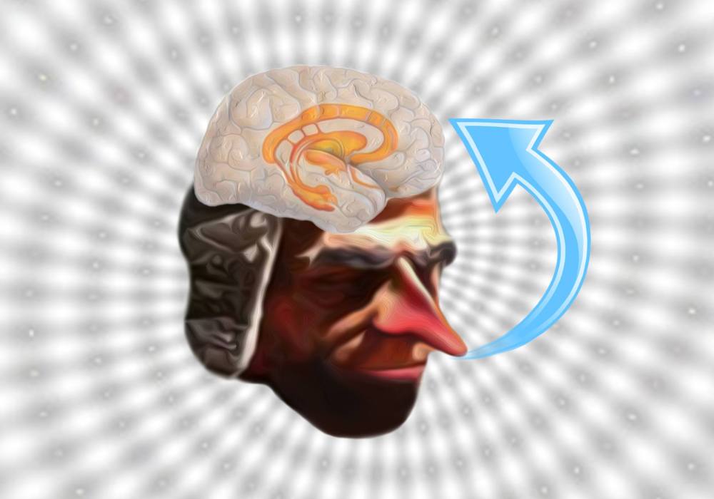
Smells, learning and memory

After seeing this article’s title, you might have thought: “That sounds rather boring. I mean, what is so interesting about the nose?” Perhaps the “memory” part aroused your curiosity, though. If that’s so, you might find the following reading worth having a look at, as you could discover some surprising things about the “nose”.

I’d like to begin by emphasising something important: we don’t actually smell with our noses; it’s the brain that identifies different odours through the central olfactory pathways, but we’ll get to that soon. What does happen in our nasal cavity is the activation of the olfactory receptors (a type of neurone) of the primary olfactory system, by chemical stimuli called odourants. The binding of odourants to the olfactory receptors’ cilia triggers the transduction process, which involves G-protein stimulation, formation of the cyclic AMP (cAMP) and membrane depolarisation, by the opening of ion channels (calcium, sodium and chloride). This complex signalling cascade results in a receptor potential which is then coded as an action potential (provided the receptor potential reaches a certain threshold) and then transmitted further along the receptor’s axons (remember, they are actually neurones!) The axons form the olfactory nerve, but they also group in small clusters and converge onto the two olfactory bulbs, in spherical structures known as glomeruli. Here, the axons synapse upon second-order neurones which form the olfactory tracts and finally project to the olfactory cortex (involved in the perception of smell) and to some structures in the temporal lobes, the medial dorsal nucleus (in the thalamus) and the orbitofrontal cortex. The last two are thought to play an important role in the conscious perception of smells. A pretty intricate process, isn’t it? But it is a lot more to olfaction than this!

Running parallel to the primary olfactory system is the accessory olfactory system. This has been shown to detect our favourite smelling chemicals, the pheromones. As I am sure most of you are aware of, pheromones are involved in reproductive behaviours, identifying individuals, aggression and submission recognition. Not only the type of chemical stimuli, but also the structures in the accessory olfactory system are different: the vomeronasal organ in the nasal cavity, the accessory olfactory bulb and last but not least the hypothalamus and amygdala (and hippocampus) as the final axonal targets. The amygdala and hippocampus are known for their implications in emotions and long-term memories (check out article about memory). Thus, olfaction also plays an important role in the integration of different odours in emotion processes, as well as explicit memory and associative meanings to odours.

Interestingly, each receptor cell is defined by only one receptor protein, which is encoded by a single receptor gene. These genes form the largest family of mammalian genes: 1000 in rodents, 350 in humans. The receptor cells have unique structures and are divided into different types according to their sensitivity to odours: each receptor type is activated by a single odour; nevertheless, one odour can activate many receptor types and the combination as well as the frequency, rhythmicity and temporal pattern of receptor stimulations encode for odour information.

Studies in Drosophila have shown another very important function of the olfactory processes: the associative learning. Gustatory unconditional and odour conditional signals both converge on the antennal lobe and mushroom body of the Drosophila, establishing learning efficacy of appetitive and aversive memories in classical conditioning. The release of certain catecholaminergic neurotransmitters such as dopamine and octopamine (the insect analogue of noradrenaline) are involved in the aversive and appetitive behaviours, respectively. In an incredibly revealing study, Lee Chi-Yu and his colleagues developed this topic in much more detail. I strongly recommend you have a look at it here.

As you might have guessed, given the fact that olfaction has a wide range of implications, its impairments are present in different mental diseases. Olfaction deficits or absence (anosmia) have been identified in Alzheimer’s disease and dementia, whereas olfactory hallucinations and weird smells are one of the main symptoms of schizophrenia.

Hopefully, you didn’t find this article too long or confusing. Did you find out new information about olfaction? If you have any questions or comments, please feel free to upload your posts. I’m looking forward to them as always.

For further information:

Bear et al., “Neuroscience”, third edition, Lippincott Williams & Wilkins

My friend’s article

Another very relevant article

Photo by Isuru Priyaranga 

Spatial memory, grid cells and…honeycomb?!

Remember when we talked about memory? One very important aspect you should never forget is that there are many types of memory (stored in different areas of the brain). Today I’d like to discuss a particular and very important type of memory, known as spatial memory. As a matter of fact, I’m going to be even more specific: this article deals with one of the several types of cells involved in spatial memory, which were recently discovered. These were called grid cells (the name is very suggestive, as you’ll see in a few moments).

You may wonder why I chose this topic. Obviously, it is of great interest for many scientists, but to be honest, my own subjectivity played a role in here as well. For those who haven’t met me, you should know this: there is no one in the world less capable of spatial self-orientation than myself! Therefore, when I heard that a young Norwegian couple (the Mosers) won the Noble Prize in 2014 for discovering some neurones specialised in location memory, it came as no surprise that I became extremely curious about their research.

As I mentioned before, grid cells are not the only neurons involved in spatial memory. In the 1970, John O’Keefe discovered navigation in the brain along with the cells that generate this process, known as place cells. These neurons are located in the hippocampus (a structure of the temporal lobe associated with long-term and spatial memory) and appear to code for location, by creating a spatial map of the environment. They are dependent on the motion’s speed and direction.

Grid cells represent a type of place cells, although unlike place cells, they are predominantly found in the medial entorhinal cortex. Some grid cells were also found in the prefrontal cortex, which is involved in forming episodic memory. A quite peculiar characteristic about these neurons is that they fire whenever the subject is at certain locations and these signalling locations form a hexagonal pattern, very similar to a honeycomb (which explains the rather odd image I chose for this article).

This honeycomb-like pattern is not only mathematically precise, but it is also very efficient as it uses a minimum number of grid cells to achieve the highest-possible resolution, thus saving energy.  It was also discovered that the firing patters remain constant during the subject’s movement, regardless its speed or direction. Moreover, unlike the response of place cells, which is based on visual stimuli, the grid cells’ firing is maintained even in the absence of sensory stimuli, thus showing an algorithm based on self-motion. In an interview for Deutsche Welle Magazine, Edvard Moser says: “It is thought that these [grid cells] are part of an internal map that is based on our own movement, so that these cells signal the distances we move and the directions we take”.

The discovery raises scientists’ hopes up in treating neurodegenerative diseases linked to memory, such as Alzheimer’s disease, as it has been demonstrated that the first symptoms of dementia appear in the entorhinal cortex. At the same time, for other people, like me, grid cells may offer an explanation as to why they still can’t find their best friend’s house, after having been there several times.

Articles related to the subject:

 Nature

 New Scientist 

 Deutsche Welle 

Image created by Isuru Priyaranga

Let’s not forget about memory!

In the previous article we talked about empathy, as the capacity to put ourselves in someone else’s shoes and understand them. We discussed about the structures involved, in a quite simplistic manner, but as it turns out things are not that simple. What makes the brain so fascinating (and hard to study as well) is the fact that each and every process, from the basic to the more advanced ones (like reasoning, planning, decision-making) require the integration of many different structures and other processes, that in turn depend on other processes and so on. A bit confusing, isn’t it? Don’t worry! If you feel lost, then you’re actually on the right track.  

Mirror neurons represent one of the basic keys behind empathy (as presented in the previous article), but they don’t explain the whole picture, though. In order for us to be able to relate to a complete stranger’s situation and feel what he’s feeling, we first need to be able to relate to ourselves. More specifically, our brain should be capable of retrieving something from our own past that is similar or triggers similar emotions to what the stranger is experiencing. (This is, of course, involuntarily and subconsciously.)

That’s where memories come in. Memory is a very common term and almost all the people know what it is about. Or do they? In fact, “memory” is quite ambiguous and imprecise; neuroscientists discuss about “types of memory”. Indeed, there are several categories in which memories are classified (visual-spatial memory, auditory memory, emotional memories, long-term memory, short-term memory – if we focus on duration – and so on). 

To spare us of too much confusion, scientists decided to divide memory into two broad categories: declarative (explicit) and non-declarative (implicit) memory. The first one refers to memories for facts and events and is the conscious component, whereas the second one includes the types of memory that don’t have a conscious component, such as memories for skills and habits, priming, classical conditioning, simple forms of associative learning, as well as simple forms of non-associative learning (habituation and sensitization).

A lot of information to take in, I know. But if you’ve manage to get this far, I promise you won’t be disappointed! The big question that has given scientists a hard time for years was: “Where exactly is memory stored?” Initially it was thought that memory is somehow spread throughout the whole brain. Well…it’s not! And we now know this thanks to one of the most famous patients in the history of Neuroscience, Henri Molaison or H.M., as he is referred to in literature.

H.M. had been suffering from epilepsy his entire life and in 1953 he underwent lobotomy to treat his seizures. Thus, he had a part of the brain removed, which included both his left and right temporal lobes. After the surgery, the epilepsy was gone, but instead something else appeared to have taken its place… H.M. was no longer able to form new memories (anterograde amnesia). To him, every day was like a new beginning: everything he did or learnt, all the people he met the day before, were completely new the day after. At least, he never got bored (which allowed neuroscientists to study this patient his entire life, as he couldn’t recall any of the tests he participated in). 

Interestingly, his old memories (from childhood, for instance) were unaffected and he was even able to learn new things, although he had no recollection o them. Thus, scientists concluded that the temporal lobes and especially the hippocampus were responsible for the formation of long-term declarative memories, but instead this type of memory was stored somewhere else (in the frontal cortex). Also, a different region of the brain is involved with memories for skills and habits (the striatum). Hence, there are specific cortical areas where memories are encoded.

This article is just to give you a bit of an understanding of how the brain deals with basically sorting our entire lives into distinct, separate “folders”, but it’s not even close to covering this whole vast subject. At some point, you might come across another article about a special type of memory that was recently discovered. 

Did memory manage to arouse your curiosity? If so, I’m looking forward to comments.  

P.S. I would like to give very special thanks to Sorin Hornoiu and Isuru Pryiaranga, who created the images for the first article and this one, respectively. I try as much as possible to use original pictures (not downloaded from the internet) and their creative skills really come in handy.