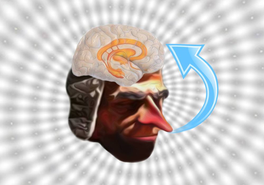
Oxytocin and Social Bonding

While most of us would be able to describe what being affectively close to someone feels like, we might find it harder to explain why and how such a connection forms.

Why do we love and what makes us love certain people? Why is love so different depending on the subject of our affection? Is it possible to measure love? What does the complete absence of love in an individual reveal about their health state? With so many questions having been formulated throughout centuries, no wonder love has become a universal conundrum. Traversing various disciplines, it not only represents the realm of the literary, but it has increasingly become one of the central focuses in philosophy, biology, social sciences and neuroscience.

As far as the neuroscientific approaches to love go, this concept is represented by affiliative bonds. Therefore, from now on we shall refer to love as such. For the sake of the reader’s personal interest, we shall further discuss affiliative interactions as they appear and manifest in humans. Affiliation describes the ability of an individual to form close interpersonal bonds with other individuals of the same species. Three prototypes of affiliation have been identified: parental (between children and their parents), pair (between romantic partners) and filial (between friends).

This article is intended to introduce the reader to the evolutionary significance and neurochemical mechanisms underlying social bonding/affiliation. As such, the above-mentioned types of affiliative behaviours will be only in part separately discussed. Instead, we shall focus on what these categories share in common, particularly, the hormone-neurotransmitter oxytocin and the concept of synchrony.

Synchrony refers to the process by which the members of a social group collaborate with each other, in order to achieve a social goal. This kind of collaboration involves concordance in time between members, at the level of behaviour and physiological processes (e.g. hormonal release, neural firing). Through these synchronous processes underlying social reciprocity, each member is introduced to the social milieu, becomes adapted to his/her environment and learns how to survive.

Intimate reciprocal relationships between two individuals in a social group help shape the individual’s moral, empathic and pro-social orientation, as well as social adaptation and self-regulation. The interaction between mother and infant is critical to the social maturation and well-being of the young. Human mothers, just like other mammals, exhibit specific postpartum behaviours, such as affectionate touch, high-pitched vocalisations, expressing positive affect, which lead to the notoriously strong mother-infant bond.

This type of specific attachment relationship coordinates the physiology of the infant with the behaviours of the mother. Moreover, this mother-infant synchrony enables the temporal alignment of the infant’s inner state with the responses of the social environment (via the mother). The absence of a proper interaction between mother and child, especially within the critical period (between 3 and 9 months after birth), has been shown to contribute to the development of autism spectrum disorders (for more information on autism, check out this previous article – Decoding autism).

Romantic attachment is another type of social bonding in humans, with significant implications to the normal psychological functioning of the individual. According to recent studies, both parental and romantic relationships share similar behavioural characteristics (gaze, touch, affects, vocalisations and coordination of these behaviours between the members of the pair) and rely on similar neuroendocrine mechanisms. These mechanisms mainly involve a nine amino-acid neuropeptide known as oxytocin.

Oxytocin acts as both a hormone and a neurotransmitter. It is associated with a variety of functions including the initiation of uterine contractions during parturition, homeostatic, appetitive and reward processes, and last but certainly not least, the formation of affiliative bonds. For the latter, oxytocin plays a very important role in social recognition, maternal behaviour and development of partner preferences.

Oxytocin is produced in the hypothalamus, by the magnocellular neurones clustered in two types of nuclei: the supraoptic and paraventricular. These neurones send projections to the posterior pituitary gland, thus engaging the oxytocin system with the hypothalamic-pituitary-adrenal axis, mediating the stress response, as well as parturition, lactation and milk ejection. Other projections from the paraventricular nucleus go to various forebrain limbic structures (e.g. amygdala, hippocampus), brainstem (e.g. ventral tegmental area) and spinal cord. There are also other areas, apart from the brain and spinal cord, which receive oxytocin signalling, such as the heart, gastrointestinal tract, uterus, placenta, testes etc. With such extensive projections, it comes as no surprise that oxytocin is involved in a wide variety of processes.

In romantic and parental attachment, oxytocin induces the motivation to initiate sexual behaviour, the formation of sexual preferences and the increased stimulant value of the infant for its mother, via its connectivity with the mesolimbic dopaminergic neurones. The neurotransmitter dopamine plays a major role in the reward-motivated behaviour. Therefore, the oxytocin-dopamine interaction is key to the motivation to bond between members of romantic or child-parent relationships.

If you were wondering why the parental attachment has so far been presented only from the perspective of the mother-child relationship, that is because in males a different hormone mediates parental behaviour. Vasopressin can be seen as the male equivalent of oxytocin, as it modulates affiliation, aggression, juvenile recognition, partner preference and parental behaviour in males. Having said that, there are studies which show that oxytocin also supports paternal behaviour and is linked to the father-typical affiliative behaviour.

Oxytocin is also very important in establishing close connection with our best friends (what is known as filial attachment). According to research in this area, children start showing selective attachment to a ‘best friend’ around the age of 3. This kind of interpersonal interaction represents the first attachment to non-kin members of society, therefore, a crucial step in the normal development of any human being.

Depending on the level of synchronous parenting children experienced during infancy, their interactions with best friends can vary in the degree of reciprocity, emotional involvement and concern for the friend’s needs. These behaviours are modulated by oxytocin. During the first 3 years of life, oxytocin secretion in humans depends on the parent’s postpartum behaviour (which is predicted by the parents’ own levels of oxytocin) and, in turn, determines the degree of empathy between close friends. Therefore, a reasonable assumption, which has been recently proven, is that children benefiting from high parental reciprocity during infancy develop better social adaptation, are more friendly and cooperative, and show greater empathy.

All in all, the social bonds we form with members of our social group, be they our family, romantic partners or friends, are dependent on certain hormones and behaviours occurring at critical stages of development. Close attachment bonds with our parents, during early infancy, are later translated into affiliations to non-kin members of the social groups, who we come across during childhood, evolving into intimate friendships during adolescence, which eventually shape the ability of the adult human to form and maintain romantic connections and provide nurture for the next generation.

What we have just discussed is of importance for different aspects. Focusing on oxytocin and synchrony provides better understanding of neurodevelopmental disorders such as autism. At the same time, this focus offers some answers to questions regarding the reasons and mechanisms underlying the many types of love us humans experience throughout our lives.

References

Feldman, R. (2012). Oxytocin and social affiliation in humans. Hormones and Behavior, 61(3),  380-391. 

Hammock, E. A. ., & Young, L. J. (2006) Oxytocin, vasopressin and pair bonding: implications for autism. Philosophical Transactions of the Royal Society B: Biological Sciences, 361(1476), 2187–2198. 

Fear and the amygdala

What is fear? Why are we sometimes afraid? Can fear be inhibited? What produces fear – the brain or the heart?

It is definitely the brain! More exactly, something in the brain – a tiny, almond-shaped structure, which sits anteriorly to the hippocampus, called the amygdala. This small part of our brain is to blame for  the perception of fearful stimuli and the physiological responses (increased heart rate, electrodermal responses etc.) to fearful stimuli.

As part of the FEAR system, the amygdala connects to the medial hypothalamus and the dorsal periaqueductal grey matter (in the midbrain), which is important in pain modulation in the dorsal horn of the spinal cord, as well as to sensory and association cortices. The lateral nucleus of the amygdala receives inputs from different brain regions, thus allowing the formation of associations, required for aversive conditioning. Following the processing in the lateral nucleus, information about the stimulus is, then, projected to the central nucleus of the amygdala, where an appropriate response to the stimuli is initiated, provided that the stimuli are detected as threatening or potentially dangerous.

The amygdala is involved in emotional learning and memory, modulating implicit learning, explicit memory, attention, social responses, emotion inhibition and vigilance.

You can find the article on memory here, to brush up a bit.

  • Implicit memory is a type of learning, which cannot be voluntarily reported or remembered. It includes the memories for skills and habits, for procedural knowledge, grammar and languages, priming, simple forms of associative learning and classical conditioning. The latter is particularly important for fear. It involved a conditioned stimulus (CS) and an unconditioned, painful/fearful stimulus (US), with US preceding CS and determining a fearful response to CS. This type of fear learning is adaptive and is known as sensitisation or acquisition.

There are two different pathways in the amygdala, important for fear conditioning. The “low road” pathway: sensory information projects to the thalamus, which directly communicates with the amygdala; this pathway is fast, modulating rapid responses of the amygdala to different types of fearful stimuli. The “high road” pathway is an indirect pathway: sensory information projects to the thalamus and from there, it is conveyed to the sensory cortex for a finer analysis; the sensory cortex, then, communicates the processed information to the amygdala. This pathway ensures that the sensory stimulus is the conditioned stimulus. So the responses of amygdala to threatening stimuli are both rapid and sure.

  • Explicit memory refers to the memory of facts and events; in the case of fear is means the processing and retention for a long time, of emotional events and information. For this type of fear learning, the amygdala interacts with the hippocampus. There is a distinction between the formation of memory for aversive experience (fear conditioning), which is based on previous experience, and explicit learning (in the hippocampus), which involves learning and remembering aversive properties of different threatening stimuli. The memory in the hippocampus is enhanced by arousal produced in the amygdala. The activation of the amygdala can make different cortical areas, not just the hippocampus, more receptive to stimuli that are adaptively important, thus ensuring that unattended, but important stimuli gain access to consciousness.
  • Social responses involve the ability to recognise a stimulus as good, bad, neutral or arousing. This ability, however, does not depend on the amygdala. There is one exception here, otherwise we wouldn’t be talking about it in the context of fear mediated by amygdala – fearful facial expressions. According to Darwin, social species, like humans, use facial expressions to detect internal emotional states of other members of the group. This function, mediated by the amygdala, is crucial in the emotional regulation of human social behaviour. Damage to amygdala has been demonstrated to result in impairment of the patient to identify fearful faces correctly and, therefore, react to them, accordingly. It should also be noted there is no need for the subject’s awareness of the fearful stimulus, for the amygdala to respond. In other words, when a fearful facial expression is presented subliminally, the amygdala will still show activation.
  • Inhibition of fear – it is actually very difficult to escape your own fears, as fear proves to be resistant to voluntary control. However, there is a process called extinction, a method of classical conditioning, where a CS previously associated with an aversive US in presented alone for a number of times, until the CS no longer signals the fearful stimulus. If the US is presented again, after the passage of time, it will evoke fear responses, but in different brain areas. So, the learned fear has been retained in memory, but extinction learning eliminates the response to fear. This mechanism of extinction relies on the activity of NMDA glutamate (excitatory) receptors in the amygdala. When these receptors are blocked, extinction is inhibited (so you will react to fearful stimuli) and when they are active, extinction is augmented (you no longer respond to aversive stimuli).
  • Vigilance – the amygdala is not necessary for the conscious experience of emotional states, but it plays a major role in increasing the vigilance of cortical response systems to emotional stimuli.

Memories about fearful events, just like other types of long-term memory, become permanent through a process of gene expression and novel protein synthesis, which is known as consolidation. Upon retrieval, the memories become susceptible to change and alteration, before they are reconsolidated, which involves additional protein synthesis.

The fact that humans (and probably other animals as well, I am sure, although not widely proven) have the ability to distinguish between emotional information and unemotional information is regarded as an evolutionary advantage. Emotional stimuli signal dangers and the ability to detect and appropriately react to them increases the chance of survival. However, exacerbated fear is detrimental to the individual who experiences it and is a sign of pathology. For instance, in atypical monopolar depression, which includes anxiety as one of the main symptoms, the amygdala is overactive and it determined lowering the threshold for emotional activation and abnormal reactions to stressful stimuli. A similar pattern can be seen as a result of partial chronic sleep deprivation or complete acute sleep deprivation.

References

 Beatty, 2001. The Human Brain – Essentials of Behavioural Neuroscience, Sage Publications Ltd., pg. 293-296

Bernard et al., 2007. Cognition, Brain and Consciousness – Introduction to Cognitive Neuroscience. 1st edition. Elsevier Ltd., pg. 373-383

Gazzaniga et al., 2002. Cognitive Neuroscience – The Biology of Mind. 2nd edition. W.W Norton & Company, Inc., pg. 553-572

Image by  Saya Lohovska. You can find her arts page here.

Consciousness – Who decides, đ˜șđ˜°đ˜¶ or your brain?

If you were to answer the question “What differentiates humans from other organisms on Earth?”, you would probably list a number of things, including the ability of humans to make “free choices” dictated by their consciousness, rather than by something organic. Am I right?

What if someone told you that this is not actually the case? I mean, what if instead of making decisions out of your own will, your brain is “deciding” for you and only after the decision has been made the brain offers you the illusion of conscious act, making you believe that you were the one who made the choice in the first place. But how is it possible that such a dichotomy exists within ourselves, between us and our own brains? Aren’t we our brains? Apparently not!

Now that I (hopefully) managed to capture your attention, I’d like to bore you a bit with some brain structure names and functions, which are necessary in order to begin to understand what’s going to come next.

The frontal lobe contains a few areas, which are involved in planning our movements, decision-making, emotions (usually associated with the decisions we are about to make), repeating previously memorised motor sequences etc. These are the areas involved in voluntary motor control, more specifically, these are the areas that make the difference between reflexes/automatisms and movements or actions we want to pursue. Moreover, all these motor areas are interconnected and also linked to areas that are part of the sensory pathways, such as the parietal , visual, somatosensory and temporal regions (which store different components of visual, auditory and somatic stimuli and are associated with many diseases, such as the inability to feel your own limbs, or recognising faces/objects etc.).

  • The primary motor cortex (PMC) is mainly involved with the execution of movements. Populations of neurones in there encode for the direction and amplitude of the movements we make, prior and during the execution.
  • The 6 pre-motor cortices, the ventral, medial (supplementary) and dorsal areas are mostly involved with planning our movements. They receive inputs from the cerebellum and basal ganglia, which play very important roles in motor learning (like acquiring new skills) and motor planning. Interestingly, different neurones in the pre-motor areas fire action potentials during execution and are inactive during planning of movements and others vice-versa, while some populations of neurones are active for both planning and execution.
  • The prefrontal cortex controls reasoning and decision-making and it is crucial for emotion as well: recall Phineas Gage’s story and how the damage to his prefrontal cortex resulted in a complete change in his personality (article here) as well as how the prefrontal cortex regulates the activity in the hypothalamus and is disrupted in major depressive disorder (article here). 
  • The limbic system (amygdala, anterior cingulate cortex, hippocampus) , which are located at the subcortical level and behind the frontal lobe, are involved with emotion, fear and the formation of memories, which are so important in our decision making. And these are just the main players, but there are many other areas, including sensory, which contribute to the planning of our actions and the choices we constantly make.

In a rather groundbreaking paper, Libet and colleagues showed that the neural processes leading to the initiation of voluntary movements begin several hundred milliseconds before the reported time of conscious intention to make the movements, as in before the subject is aware of the intention to move. They demonstrated using the readiness-potential (negative electrical potential recorded at the scalp) that brain activity involved in decision-making starts before our brains is conscious of the actions. This is also known as ‘preparatory set”.

Dick Swaab proposed that the unconscious brain areas are active before the conscious ones, in order to enable us to make decisions rapidly and effectively, as the conscious systems require time to process and analyse the pros and cons of every decision. And although it is good to consider the consequences of your actions, there are many other decisions about apparently insignificant things, which we make and need to be fast (like for example, running away from a car you see coming). In a dangerous situation, for example, the parts of the brain involved in consciousness might consider the state of your legs, how capable they are of moving fast at that point, your heart rate, blood pressure, levels of energy needed for that action
Well, by the time your brain finishes analysing all these, you will be most certainly dead.

Another interesting idea Swaab suggested was regarding the reason why we have consciousness of our actions and the things that happen to us in the first place. We need to be conscious of our own experiences so that we learn to avoid negative things in the future and also act upon things that require intervention, such as a wound that needs to be treated. Although the brain seems to be able to plan an action independently of our awareness of it, other brain areas are involved in the execution (as previously mentioned) and the communication between these different parts which fulfil different roles results in consciousness. Exactly why and how evolutionary biology has managed to make us more than just some purely mechanical creatures remains a mystery and still poses many challenges to this field of research, inviting philosophy to have its take on this matter, which many times has proved to be useful.

Swaab also wonders to what extent are criminals, pedophiles, murderers to blame for their bad actions, when it is in fact not them, but their unconsciousness/instincts that dictate them what to do. When considering that people with brain damage resulting in impaired or lack of consciousness (schizophrenia, dementia, multiple sclerosis etc.) sometimes hurt other and are not convicted, you might think that it is right to assume that all criminal acts should be tolerated. However, the difference here is that people not suffering from such disorders are aware of their actions and are capable of stopping them. Although pedophilia is considered a psychiatric disorder, unlike the neurodevelopmental and neurodegenerative ones, it can be controlled by the individual, so that the individual is able to refrain from acting according to his/her instincts. Libet and his team of researchers mention in their study that individuals, although only aware of the intention to make a particular action after the intention has been formed in their brains, are able to “abort the performance” of the action, meaning that they have a conscious “veto”.

They also emphasise the difference between spontaneous, rapidly performed actions, and actions in which a preplanning of the experience occurred (taking into account alternative choices, for instance). This second type of voluntary movements, involving conscious deliberation prior to the act, might actually rely on conscious initiation and control, rather than non-conscious commands. However, this hypothesis has not yet been proved experimentally, in a way the “unconsciousness before consciousness” one has.

So, as it turns out, most of the times we are aware of our brain’s decisions only after they have already been made, and free will seems to be an illusion.

References

Libet et al. paper

Antonio Damasio,1995. Decartes’ Error. Vintage Books, pp. 71-73

Dick Swaab, 2014. We are our brains – From the womb to Alzheimer’s. Penguin Books, pp.326-338

Image by Saya Lohovska. You can find her arts page here.

Depression and why some of us are SAD

I am warning you, this is going to be a long one! But it’s interesting, I promise.

There is a lot of confusion and mixed opinions when it comes to depression. Some people use the term inappropriately, to describe what is in fact grief (the feeling of sadness that humans, and presumably other animals, experience after a loved one has died, for instance), others tend to label depressed individuals as “weak”, “selfish”, “useless”, “cowards” etc.

At the same time, for the past 30 years, there have been extremely important discoveries in the field of affective disorders, which have helped eliminate many misconceptions and laid the foundation for a better understanding of what this set of disorders (affective disorders) are and what is actually happening in the brain of the ones “affected”. Moreover, according to some new theories, depression is in fact an evolutionary advantage in situations such as physical illness and dominance. When the body is sick it needs time and energy in order to recover, so the organism experiences depression in order to avoid activity and focus on recovery; in nature, many animals with a dominant status are forced (by a variety of factors) to occupy a lower hierarchical level, in which case “depressive” behaviours such as avoiding eye contact or sexual contact helps reduce the risk of attack by other dominant, more powerful individuals. There are many theories, as you can see, and this article is meant to present and analyse some of them.

We should start by clarifying a very important aspect: depression is different from grief. While the latter is a normal reaction to some external factor(s) with a negative emotional impact on our day-to-day lives, and dissipates by itself after a certain period of time, depression is a pathological, abnormal condition (either in its own right or as a symptom of other metabolic or neurodegenerative diseases). However, it should be noted, and this is one of the key concepts in understanding depression, that the way individuals interpret and react to various external events, which affect their mood, differs, which means that some individuals have a stringer predisposition to depression than others. Now, why is that? A variety of factors, both genetic and epigenetic (developmental, such as child abuse, neglect) play a role and often act synergistically, but we will deal with them (especially, the genetic factors) a bit later in the article.

Different types of depression

Major depressive disorder, also known as “the classical depression”, which is characterised by insomnia, anorexia and lack of joy and interest in things. At the opposite side of the spectrum, there is atypical depression, which manifests itself through increased sleepiness, weight gain and anxiety. Dysthymia is another form of depression, more difficult to diagnose, due to the fact that it presents itself with mild depressive symptoms. All these types discussed so far have been categorised as monopolar.

Bipolar depression refers to a kind of depression accompanied by periods of mania – manic episodes are characterised by elevated, euphoric mood, impulsiveness, hyperactivity and even psychotic symptoms (hallucinations, delusions). A case described by Dick Swaab in his book “We are our brains – from the womb to Alzheimer’s” portrays a woman, who developed mania following the death of her husband. She would talk and laugh hysterically, call the police in the middle of the night for no reason and eventually began to make up stories about people whom she had never met before, but who she believed were longtime friends of hers. After her manic episodes disappeared as a result of treatment, she developed severe depression. Luckily, her story has a happy ending, as she made a full recovery.

Bipolar depression is also associated with Seasonal Affective Disorder (SAD), characterised by extreme mood seasonal swings. In this article, I have dedicated an entire section to SAD, so I am not going to delve into it for now. Given all these particularities of BD, it is often regarded as a separate disorder (bipolar disorder or manic disorder), rather than another type of depression. As there are so many things to mention about depression, I will leave BD for future article.

Diagnosing depression

In order to be diagnosed with depression, one must have at least one of the two main symptoms: persistent sadness and marked loss of interest, as well as at least five secondary symptoms: disturbed sleep (either increased or decreased), disturbed appetite (increased or decreased), fatigue, poor concentration, feeling of worthlessness and excessive guilt, suicidal thoughts.

Depending on the number of these symptoms, as well as the degree to which they manifest, monopolar depression can be sub-divided into: sub-threshold depression (fewer than five secondary symptoms; no treatment needed), mild depression (fewer than five, but in excess secondary symptoms), moderate depression (more than five, plus functional impairment between mild and severe depression) and severe depression (most of the secondary symptoms and also true psychotic symptoms – yes! they can occur in severe monopolar depression as well, not just BD).

Biochemical pathways and brain systems involved in depression

In Ancient Greece, there was a biochemical theory of depression. It was believed that depression was caused by the failure of liver to eliminate toxic substances from the digested food, resulting in the accumulation of “black bile” (melan means “black” and chole means “bile”, which give the words melancholy). Biochemical theories nowadays have at their core three monoamines, which I am sure you are all familiar with: noradrenaline (a neuromodulator very similar to adrenaline) and serotonin and dopamine.

These two substances have long and diffuse projections throughout the nervous system and in levels lower than otherwise normal, they are said to be involved in affective disorders. For example, drugs such as Reserpine, used to treat the positive symptoms of schizophrenia by depleting dopamine (and also serotonin and noradrenaline) elicited depressive symptoms in schizophrenic patients.

Therapies involving monoamines

The idea is, you want to higher levels of monoamines in order to treat depression. Enzymes involved in the monoamine re-uptake mechanism from the synaptic cleft back into the presynaptic level and enzymes involved in the monoamine metabolism, such as monoamine oxidases (MAO) are the most common targets for the majority of anti-depressants.

  • Selective serotonin re-uptake inhibitors (SSRI) and selective noradrenaline re-uptake inhibitors (SNRI) – Prozac (Fluoxetine), Zoloft (Sertraline), Celexa (Citalopram), Paxil (Paroxetine) block serotonin reuptake and Effexor/Viepax/Trevilor/Lanvexin (Venlafaxine), Cymbalta (Duloxetine) inhibit the noradrenaline reuptake enzymes. For those of you who are currently under this treatment, be careful! Side-effects such as sexual dysfunction, insomnia, increased aggression and self-harm/suicide can occur. Moreover, SSRI are not so effective. They have a very long induction, which means that it takes a long time (2-3 weeks) for the therapeutic effects to start working, during which time there is a high risk of suicide (due to depression). They also have a placebo effect of 50%, which is not necessarily a bad thing as long as it works, but raises the question whether the monoamine hypotheses is really that valid in the case of depression.
  • Tricyclic antidepressants – also block the reuptake mechanism, resulting in more monoamines in the synaptic cleft. Amitril (Amitriptyline), Aventyl/Norpress/Noritren (Nortriptyline) and Tofranil (Imipramine) are a few examples. They are derived from Phenothiazines (such as Chlorpromazine), which are antipsychotic drugs (used to treat schizophrenia). Some of the side-effects are: chronic pain and suicide overdose.
  • MAO inhibitors – Nardil/Nardelzin (Phenelzine), USAN (Thanylcypromine), Marplan/Enerzer (Izocarboxazid) and Amira/Aurorix/Clobemix (Moclobemide) are very effective and widely prescribed for in major depressive disorder, bipolar disorder and anxiety disorder, although the first three pose the high risk of hypertensive crisis and death if the patient is consuming cheese or wine.

The big problem with these drug therapies is dependence – if antidepressants, especially Paroxetine and Venlafaxine are administered for a long period of time and then stopped, the patient is likely to experience Antidepressant discontinuation syndrome, characterised by flu-like symptoms, motor and cognitive disturbances.

Non-drug therapies

An alternative to pharmaceutical treatments is represented by transcranial magnetic stimulation (TSM) of the cortex, electroshock therapy – this is, apparently, very effective, BUT might result in impaired memory – and gene therapies. The latter refers to the insertion, via a vector or a plasmid, of genes that encode neurotransmitter molecules, receptor proteins or neurotrophic and neuroprotective substances. Given that many variations in genes for chemical messengers in the brain are responsible for the predisposition of certain individuals to depression, gene therapies, although still at a developing stage, provide powerful approaches to the treatment of affective disorders.

Over-activation of the stress axis

Another theory for the development of depression, which goes hand-in-hand with the “monoamine hypothesis” is that in depressed individuals there is an exaggerate amount of cortisol (a steroid) in the blood, which can affect the brain. Basically, our brains react to stressful situations by producing some hormones in the hypothalamus and pituitary gland (hypophysis), which eventually result in the production of cortisol. In turn, cortisol acts on these structures to inhibits their activity and, thus, preventing further increases in its level – this is an example of a negative feedback mechanism.

In normal people, a stressful situation will result in increased levels of cortisol, but this steroid will then revert to its normal levels. In depressed individuals, the stress axis (hypothalamus-pituitary-adrenal axis) becomes hyperactive and, as a result, a stressful event will result in the overproduction of cortisol.

In excess, cortisol affects brain structures involved in the control of emotions and fear, such as the cingulate cortex and amygdala (which explains the anxiety symptoms experienced by people suffering from atypical depression) and memory, such as the hippocampus, which explains the cognitive dysfunctions. Moreover, the activity in the prefrontal cortex, which normally inhibits the hypothalamus (overactive in depression) is decreased by cortisol. So, really, it is like a vicious circle.

Why is the stress axis hyperactive in the first place? Possibly due to decreased sensitivity of the cortisol receptors to cortisol, which might be the result of genetic as well as developmental factors (previously mentioned).

Monoamines play a role here, as increased levels of monoamines (by the administration of antidepressants) can determine neurogenesis in the prefrontal cortex and hippocampus, so these areas can function properly again and can, thus, inhibit the hypothalamus, so no longer hyperactivity of the stress axis!

Seasonal affective disorder (SAD)

Although I am planning to write about bipolar disorders in another article, I thought it is worth discussing SAD in this article as well, given that so many people, especially those living in the Northern hemisphere, suffer from it.

In the References section there is a document called “The recent history of seasonal affective disorder (SAD)”, which is a transcript of the 2013 Witness Seminar in London. I highly recommend this reading for two reasons: it is full of remarkable, extremely important information regarding SAD and the participants at this seminar included personalities such as Prof. Josephine Arendt, Prof, Norman Rosenthal, Prof. Alfred Lewy, Prof. Rob Lucas, who are pioneers of the SAD diagnostic criteria and underlying causes (for instance, Rosenthal is the first psychiatrist who diagnosed SAD).

As many of you probably know, and sadly from personal experience, SAD is a seasonal mood change disorder, a type of bipolar disorder, which determines depression during the autumn/winter seasons and hypomania during summer. In order to understand SAD, we must remember a few things about the circadian rhythm, which I have previously discussed in two articles: Why “sleep” and Even flies sleep and learn. In short, we have an internal, genetic “clock” inside our brains (in the Suprachiasmatic nucleus – SCN), which determines the body to function in an approximately 24-hour cycle and which is also entrained by the light-dark cycle. This is not only a circadian (day-night) clock, but also a seasonal clock, which means that changes in the environment (especially light and temperature) across the year entrain this clock and determine physiological and psychological changes in our bodies.

In SAD, there is an abnormal secretion of melatonin (the hormone that triggers sleep, when it is dark outside). Light inhibits this hormone: cells in our retina, which are not coding for visual information, send projections via a distinct pathway than the rods and cones. These cells, containing  the peptide melanOPSIN, project via the retinohypothalamic tract to the SCN, “telling” the brain that it is dark outside, so the brain (SCN) determines the synthesis and release of melatonin from the pineal gland. When there is light outside, the production of melatonin is inhibited. The duration of melatonin secretion is also affected by the circannual changes – long secretion in short days and short in long days. The scientists who took part in the Witness Seminar discovered that melatonine production was increased during the depressive/winter phase and that sunlight decreased its production, thus, alleviating the symptoms of depression in SAD. A note here, sunlight is an effective treatment for SAD, not ordinary room light. This explains why, during winter, when people tend to spend more time indoors, their levels of melatonin increase. The reasons why room light does not inhibit melatonin production are the intensity of light (sunlight is five times more intense than room light) and spectral differences. More about SAD and bipolar disorder in a future article!

I hope this article made sense and that you enjoyed reading it!

References
SAD – Pdf of The Witness Seminar transcript

Beatty, 2000. The Human Brain – Essentials of Behavioural Neuroscience. Sage Publications. Inc., pg.464-471

Dick Swaab, 2014. We are our brains – From the womb to Alzheimer’s. Penguin Books, pp. 112-122

Image by Damaris Pop

Decoding autism

About a year ago, I posted an article entitled Autism, which was meant to be more of an introduction into autistic spectrum disorders. Since then, I’ve been meaning to come back to this topic and provide more details about the mechanisms leading to autism, but I knew I need to do some proper research, which my student schedule didn’t really allow at that point.

The reason why I didn’t post any articles in the past three months is mainly my uni dissertation, which took up a lot of time. My chosen topic was Cellular mechanisms of autistic spectrum disorders. This assignment offered me the chance to read a lot of scientific papers and have a far better understanding of autism than I had before. As you probably guessed, this article draws quite a lot on my dissertation.

Some clarifications

When we refer to autism, we must be well aware that this is just an extreme end of the spectrum and that different neurodevelopmental disorders, sharing particular symptoms, are grouped under the term autistic spectrum disorders (ASD). The symptoms that characterise ASD are: impairments in social interaction, communication deficits and repetitive behaviours. Several factors have been linked to ASD, including gene dysregulations, alterations of the immune system and even environmental risk factors.

Discussing all the possible causes of ASD, could probably fit in a book, rather than a blog article, so I will only focus on gene dysregulations. However, if you have any kind of questions, feel free to post them in the comment section and I will try my best to answer them.

About glutamate and one its receptors

In biochemistry there are 20 different amino acids (the building blocks of proteins) and one of them is glutamate. This particular amino acid is very important because, apart from its role in the formation of proteins, it is also the main excitatory neurotransmitter in the brain. This means that glutamate is used to help the neurons communicate with each other and give rise to all sorts of brain activities we know about, including learning and memory.

When two neurons interact at the synapse (the space between the terminals of two interacting neurons), the neurotransmitter is released from the first neuron’s terminal and comes in contact with the second neuron’s terminal. But here’s the trick: in order for the neurotransmitter to have an effect on the second neuron, it has to activate certain structures on its terminal, called receptors. In the case of glutamate, there are three types of receptors, involved in different functions and with different mechanisms. The one we will be focusing on is the type I metabotropic glutamate receptor (mGluR). When this receptor interacts with glutamate, it leads to the translation of specific proteins involved in a process known as long-term depression (LTD), which influences learning and memory. Increased LTD is thought to play a key role in the development of ASD.

The FMRP protein

A few genes have been found to regulate the activity of mGluR and, through it, several cognitive processes. The Fragile X Mental Retardation 1 (FMR1) gene, which codes for the FMRP protein, is one of these genes. It interacts with mGluR-dependent proteins and is thought to regulate synaptic plasticity. During embryonic development, FMRP plays an important role in neural differentiation. Therefore, a mutation in the FMR1 gene leading to the absence of FMRP (a loss-of-function mutation) results in restricted brain development, impaired cognitive functions and autistic symptoms (previously mentioned). This mutation has been found in patients suffering from autism and especially from another brain disease, Fragile X Sydrome, which is the most common monogenic (determined by a mutation in a single gene) cause of autism. The activity of FMRP suppresses mGluR-dependent LTD, by inhibiting the synthesis of proteins involved in this process. Therefore, the absence of FMRP results in LTD, which primarily leads to mental disability.

Neuroligins and Neurexins

These represent transgenic protein families, which mediate synapse maturation in neurons using glutamate. Mutations affecting members of neuroligin and neurexin families have been found to be associated with autistic-like behaviours. The effects of these mutations on the brain are very specific, affecting cells in brain regions involved in learning and memory (CA1 pyramidal cells of the hippocampus, the Purkinje cells of the cerebellum), language (brainstem) and social interaction (somatosensory cortex). In normal situations, neuroligins and neurexins trigger mGluR-induced LTD, but their translation is inhibited by FMRP. The absence of FMRP leads to the loss of this inhibition, which results in mGluR-induced LTD.

Possible therapeutic strategies

Research on the links between mGluR transmission and genes involved in neural development resulted in a variety of therapeutic strategies for ASD. Genetic mGluR reduction and mGluR antagonists (drugs that act on this receptor by preventing glutamate to activate it) are the most common examples of treatments. Potential therapeutic candidates are Fenobam and Lithium, which act as antagonists at mGluR. Administered on animal models and on humans suffering from Fragile X Syndrome, these drugs have resulted in cognitive and behavioural improvements. Although there is hope in treating ASD, there is still a lot of research that needs to be done, especially since even diagnosing different autistic spectrum disorders is still a challenging task.

I hope this article gave you a little more insight into the mechanisms underlying autism and that you enjoyed reading it. As I mentioned above, any questions about this topic are welcomed.

References:

Baudouin, S. J. (2014). Heterogeneity and convergence: the synaptic pathophysiology of autism. European Journal of Neuroscience, 39(7), 1107-1113.

Berry-Kravis, E., Hessl, D., Coffey, S., Hervey, C., Schneider, A., Yuhas, J., Hutchison, J., Snape, M., Tranfaglia, M., Nguyen, D. V. & Hagerman, R. (2009). A pilot open label, single dose trial of fenobam in adults with fragile X syndrome. Journal of Medical Genetics, 46(4), 266-271.

Espinosa, F., Xuan, Z., Liu, S. & Powell, C. M. (2015). Neuroligin 1 modulates striatal glutamatergic neurotransmission in a pathway and NMDAR subunit-specific manner. Frontiers in synaptic neuroscience, 7, 11-11.

Etherton, M., Foeldy, C., Sharma, M., Tabuchi, K., Liu, X., Shamloo, M., Malenka, R. C. & Suedhof, T. C. (2011). Autism-linked neuroligin-3 R451C mutation differentially alters hippocampal and cortical synaptic function. Proceedings of the National Academy of Sciences of the United States of America, 108(33), 13764-13769.

Gao, R. & Penzes, P. (2015). Common Mechanisms of Excitatory and Inhibitory Imbalance in Schizophrenia and Autism Spectrum Disorders. Current Molecular Medicine, 15(2), 146-167.

Nosyreva, E. D. & Huber, K. M. (2006). Metabotropic receptor-dependent long-term depression persists in the absence of protein synthesis in the mouse model of fragile X syndrome. Journal of Neurophysiology, 95(5), 3291-3295.

Rojas, D. C. (2014). The role of glutamate and its receptors in autism and the use of glutamate receptor antagonists in treatment. Journal of Neural Transmission, 121(8), 891-905.

Zalfa, F. & Bagni, C. (2004). Molecular insights into mental retardation: Multiple functions for the Fragile X mental retardation protein? Current Issues in Molecular Biology, 6, 73-88.

Image by Isuru Priyaranga

Don’t be anxious about anxiety!

I remember when I was a small child and my mum or my uncle would take me out to one of my hometown’s parks or to the shopping centre. For some reason, I so often experienced an unexplainable fear and even dizziness and the terror that I might faint. I also had the feeling I couldn’t walk in a straight line. But no one noticed. Whenever I went to an indoor show or a classical music concert where people were sat on their seats and all they had to do was watch something and not move, talk or most importantly, look at me, I was fine. Little did I know what the problem was as it never occurred to me it was a problem at all. I knew I was shy and self-conscious and in my head that was the reason for my fears of crowds.

After I hit puberty, those irrational fears and the following symptoms became amplified and I started to seek for some scientific explanations. By reading and talking to different people I finally found out about agoraphobia. As the name suggests, agoraphobia is basically the fear of open and/or crowded spaces. The most important steps, I think, in dealing with an anxiety is first of all realising you have one and identifying the type.

Anxiety disorders are very common worldwide (with about 2% of the population suffering from them) and they are characterised by the pathological expression of fear. The most common types of anxieties are: agoraphobia, panic disorder, obsessive-compulsive disorder, social phobia, specific phobia, generalised phobia, post-traumatic stress disorder.The manifestations as well as the characteristics and the severity of anxiety disorders differ from person to person. Moreover, some anxieties can derive from other anxieties, like panic disorders. No wonder it took me a while to figure out what was going on with me. Here’s the thing and I would like people who suffer or have suffered from anxiety disorders to think about it: we often do not realise we have an anxiety (because we believe the causes underling the symptoms are different, like lack of self-confidence, heart attacks, pure coincidence etc.) or we just refuse to admit the reality.

Although anxiety has been mentioned in scientific literature since the 16th century, it wasn’t until the 1800s when it started to be considered  a mental illness. Before that, people attributed physiological and hormonal causes to anxieties.

Modern medical advances like fMRI and PET have made possible the discovery of the major role of the hypothalamic-pituitary-adrenal (HPA) axis in anxiety formation and development. Through a cascade of hormones released by this three-structure system, the brain responds to stress by activating the adrenal glands to produce cortisol. This, in turn, determines physiological changes which lead to exaggerated fight-or-flight reactions.

We shouldn’t pin all the blame on the hypothalamus though, as it only obeys two other structures: the amygdala and the hippocampus (which respond to the information processed in the neocortex). In this case, the amygdala and the hippocampus act as antagonists – the amygdala has a positive effect on the activation of the HPA axis, whereas the hippocampus suppressed this activation. This is how the normal fight-or-flight responses are regulated. Nevertheless, in patients suffering from anxiety disorders, hippocampal damage due to continuous exposure to cortisol (probably as a result of amygdala hyperactivity) leads to more cortisol being resealed from the adrenal medulla, thus the symptoms of anxiety becoming even more pronounced.

Several treatments, ranging from anxiolytic medications (benzodiazepines, alcohol, serotonin-selective reuptake inhibitors etc.) to psychotherapy have been developed in order to heal anxieties. Psychotherapy aims to get the patient accustomed to the stressor (the stimulus that produces anxiety) and, at the same time, to assure them of the extremely low risks potentially posed by that stimulus. In time, the fear of the stressor would disappear as the neuronal connections involving the stimulus processing would be altered.

I know I put between brackets alcohol as one of the many treatments against anxiety disorders. Indeed, due to its stimulating effects on the main inhibitory neurotransmitter, GABA. Essentially all drugs that can activate this neurotransmitter are considered anxiolytic, meaning they are able to treat anxieties. Keep in mind, though: This is should not be an excuse for people to become alcoholics 😛

In my case, the anxiety went away by itself, or maybe it was just me who kept on going to crowd places and telling to myself nothing bad was ever going to happen; which, to be honest, is a bit unrealistic – bad things can actually happen, but we should try to prevent them, instead of fearing them to the point when we would refuse to leave the house.

Hopefully, this article gave you a clearer idea about what triggers anxiety disorders and also made the anxious ones more confident that their fears don’t have to last forever.

Further information:

Article about anxiety

Short video on anxiety 

Documentary about anxiety

Bear et al., 2006. Neuroscience – Exploring the Brain. s.l.:Lippincott Williams & Wilkins pp. 665-670

Picture by Damaris Pop

Smells, learning and memory

After seeing this article’s title, you might have thought: “That sounds rather boring. I mean, what is so interesting about the nose?” Perhaps the “memory” part aroused your curiosity, though. If that’s so, you might find the following reading worth having a look at, as you could discover some surprising things about the “nose”.

I’d like to begin by emphasising something important: we don’t actually smell with our noses; it’s the brain that identifies different odours through the central olfactory pathways, but we’ll get to that soon. What does happen in our nasal cavity is the activation of the olfactory receptors (a type of neurone) of the primary olfactory system, by chemical stimuli called odourants. The binding of odourants to the olfactory receptors’ cilia triggers the transduction process, which involves G-protein stimulation, formation of the cyclic AMP (cAMP) and membrane depolarisation, by the opening of ion channels (calcium, sodium and chloride). This complex signalling cascade results in a receptor potential which is then coded as an action potential (provided the receptor potential reaches a certain threshold) and then transmitted further along the receptor’s axons (remember, they are actually neurones!) The axons form the olfactory nerve, but they also group in small clusters and converge onto the two olfactory bulbs, in spherical structures known as glomeruli. Here, the axons synapse upon second-order neurones which form the olfactory tracts and finally project to the olfactory cortex (involved in the perception of smell) and to some structures in the temporal lobes, the medial dorsal nucleus (in the thalamus) and the orbitofrontal cortex. The last two are thought to play an important role in the conscious perception of smells. A pretty intricate process, isn’t it? But it is a lot more to olfaction than this!

Running parallel to the primary olfactory system is the accessory olfactory system. This has been shown to detect our favourite smelling chemicals, the pheromones. As I am sure most of you are aware of, pheromones are involved in reproductive behaviours, identifying individuals, aggression and submission recognition. Not only the type of chemical stimuli, but also the structures in the accessory olfactory system are different: the vomeronasal organ in the nasal cavity, the accessory olfactory bulb and last but not least the hypothalamus and amygdala (and hippocampus) as the final axonal targets. The amygdala and hippocampus are known for their implications in emotions and long-term memories (check out article about memory). Thus, olfaction also plays an important role in the integration of different odours in emotion processes, as well as explicit memory and associative meanings to odours.

Interestingly, each receptor cell is defined by only one receptor protein, which is encoded by a single receptor gene. These genes form the largest family of mammalian genes: 1000 in rodents, 350 in humans. The receptor cells have unique structures and are divided into different types according to their sensitivity to odours: each receptor type is activated by a single odour; nevertheless, one odour can activate many receptor types and the combination as well as the frequency, rhythmicity and temporal pattern of receptor stimulations encode for odour information.

Studies in Drosophila have shown another very important function of the olfactory processes: the associative learning. Gustatory unconditional and odour conditional signals both converge on the antennal lobe and mushroom body of the Drosophila, establishing learning efficacy of appetitive and aversive memories in classical conditioning. The release of certain catecholaminergic neurotransmitters such as dopamine and octopamine (the insect analogue of noradrenaline) are involved in the aversive and appetitive behaviours, respectively. In an incredibly revealing study, Lee Chi-Yu and his colleagues developed this topic in much more detail. I strongly recommend you have a look at it here.

As you might have guessed, given the fact that olfaction has a wide range of implications, its impairments are present in different mental diseases. Olfaction deficits or absence (anosmia) have been identified in Alzheimer’s disease and dementia, whereas olfactory hallucinations and weird smells are one of the main symptoms of schizophrenia.

Hopefully, you didn’t find this article too long or confusing. Did you find out new information about olfaction? If you have any questions or comments, please feel free to upload your posts. I’m looking forward to them as always.

For further information:

Bear et al., “Neuroscience”, third edition, Lippincott Williams & Wilkins

My friend’s article

Another very relevant article

Photo by Isuru Priyaranga 

Spatial memory, grid cells and…honeycomb?!

Remember when we talked about memory? One very important aspect you should never forget is that there are many types of memory (stored in different areas of the brain). Today I’d like to discuss a particular and very important type of memory, known as spatial memory. As a matter of fact, I’m going to be even more specific: this article deals with one of the several types of cells involved in spatial memory, which were recently discovered. These were called grid cells (the name is very suggestive, as you’ll see in a few moments).

You may wonder why I chose this topic. Obviously, it is of great interest for many scientists, but to be honest, my own subjectivity played a role in here as well. For those who haven’t met me, you should know this: there is no one in the world less capable of spatial self-orientation than myself! Therefore, when I heard that a young Norwegian couple (the Mosers) won the Noble Prize in 2014 for discovering some neurones specialised in location memory, it came as no surprise that I became extremely curious about their research.

As I mentioned before, grid cells are not the only neurons involved in spatial memory. In the 1970, John O’Keefe discovered navigation in the brain along with the cells that generate this process, known as place cells. These neurons are located in the hippocampus (a structure of the temporal lobe associated with long-term and spatial memory) and appear to code for location, by creating a spatial map of the environment. They are dependent on the motion’s speed and direction.

Grid cells represent a type of place cells, although unlike place cells, they are predominantly found in the medial entorhinal cortex. Some grid cells were also found in the prefrontal cortex, which is involved in forming episodic memory. A quite peculiar characteristic about these neurons is that they fire whenever the subject is at certain locations and these signalling locations form a hexagonal pattern, very similar to a honeycomb (which explains the rather odd image I chose for this article).

This honeycomb-like pattern is not only mathematically precise, but it is also very efficient as it uses a minimum number of grid cells to achieve the highest-possible resolution, thus saving energy.  It was also discovered that the firing patters remain constant during the subject’s movement, regardless its speed or direction. Moreover, unlike the response of place cells, which is based on visual stimuli, the grid cells’ firing is maintained even in the absence of sensory stimuli, thus showing an algorithm based on self-motion. In an interview for Deutsche Welle Magazine, Edvard Moser says: “It is thought that these [grid cells] are part of an internal map that is based on our own movement, so that these cells signal the distances we move and the directions we take”.

The discovery raises scientists’ hopes up in treating neurodegenerative diseases linked to memory, such as Alzheimer’s disease, as it has been demonstrated that the first symptoms of dementia appear in the entorhinal cortex. At the same time, for other people, like me, grid cells may offer an explanation as to why they still can’t find their best friend’s house, after having been there several times.

Articles related to the subject:

 Nature

 New Scientist 

 Deutsche Welle 

Image created by Isuru Priyaranga

Let’s not forget about memory!

In the previous article we talked about empathy, as the capacity to put ourselves in someone else’s shoes and understand them. We discussed about the structures involved, in a quite simplistic manner, but as it turns out things are not that simple. What makes the brain so fascinating (and hard to study as well) is the fact that each and every process, from the basic to the more advanced ones (like reasoning, planning, decision-making) require the integration of many different structures and other processes, that in turn depend on other processes and so on. A bit confusing, isn’t it? Don’t worry! If you feel lost, then you’re actually on the right track.  

Mirror neurons represent one of the basic keys behind empathy (as presented in the previous article), but they don’t explain the whole picture, though. In order for us to be able to relate to a complete stranger’s situation and feel what he’s feeling, we first need to be able to relate to ourselves. More specifically, our brain should be capable of retrieving something from our own past that is similar or triggers similar emotions to what the stranger is experiencing. (This is, of course, involuntarily and subconsciously.)

That’s where memories come in. Memory is a very common term and almost all the people know what it is about. Or do they? In fact, “memory” is quite ambiguous and imprecise; neuroscientists discuss about “types of memory”. Indeed, there are several categories in which memories are classified (visual-spatial memory, auditory memory, emotional memories, long-term memory, short-term memory – if we focus on duration – and so on). 

To spare us of too much confusion, scientists decided to divide memory into two broad categories: declarative (explicit) and non-declarative (implicit) memory. The first one refers to memories for facts and events and is the conscious component, whereas the second one includes the types of memory that don’t have a conscious component, such as memories for skills and habits, priming, classical conditioning, simple forms of associative learning, as well as simple forms of non-associative learning (habituation and sensitization).

A lot of information to take in, I know. But if you’ve manage to get this far, I promise you won’t be disappointed! The big question that has given scientists a hard time for years was: “Where exactly is memory stored?” Initially it was thought that memory is somehow spread throughout the whole brain. Well…it’s not! And we now know this thanks to one of the most famous patients in the history of Neuroscience, Henri Molaison or H.M., as he is referred to in literature.

H.M. had been suffering from epilepsy his entire life and in 1953 he underwent lobotomy to treat his seizures. Thus, he had a part of the brain removed, which included both his left and right temporal lobes. After the surgery, the epilepsy was gone, but instead something else appeared to have taken its place… H.M. was no longer able to form new memories (anterograde amnesia). To him, every day was like a new beginning: everything he did or learnt, all the people he met the day before, were completely new the day after. At least, he never got bored (which allowed neuroscientists to study this patient his entire life, as he couldn’t recall any of the tests he participated in). 

Interestingly, his old memories (from childhood, for instance) were unaffected and he was even able to learn new things, although he had no recollection o them. Thus, scientists concluded that the temporal lobes and especially the hippocampus were responsible for the formation of long-term declarative memories, but instead this type of memory was stored somewhere else (in the frontal cortex). Also, a different region of the brain is involved with memories for skills and habits (the striatum). Hence, there are specific cortical areas where memories are encoded.

This article is just to give you a bit of an understanding of how the brain deals with basically sorting our entire lives into distinct, separate “folders”, but it’s not even close to covering this whole vast subject. At some point, you might come across another article about a special type of memory that was recently discovered. 

Did memory manage to arouse your curiosity? If so, I’m looking forward to comments.  

P.S. I would like to give very special thanks to Sorin Hornoiu and Isuru Pryiaranga, who created the images for the first article and this one, respectively. I try as much as possible to use original pictures (not downloaded from the internet) and their creative skills really come in handy.