On the nature of pain

Last week, a manatee was found in Florida waters, with the word ‘Trump’ scraped on its back. Although this kind of ruthless mutilation is horrific in itself, I started wondering if the animal felt any kind of pain.

I must admit, up until I came across the news about what happened to the manatee in Florida, I knew very little about manatees, in general. And my first thought was whether, during the scraping proccess, this manatee suffered at all. To my dismay, there were not many scientific papers dealing with the somatosensory system in manatees. However, I did find something that eased my soul a little bit: one of the articles reporting on the dreadful event states that the scratch was done in the algae growing on the animal’s back. Still, in the same article it is said that manatees have sensory hairs and nerves in their skin, which means that, if the cuts had touched the skin, they could have caused pain; not to mention the infection that the skin was at risk of, due to the open wounds.

The video below shows the above-mentioned manatee swimming, with the human-made scars on its back.

After reading all these news articles, I was left with some questions that kept occupying my mind: What are manatees?; To what extent can they feel pain?; And can we talk about ‘pain’ at all in manatees, or just nociception? Lastly, how did pain evolve throughout the animal kingdom?

What are mantees?

Also known as ‘sea cows’, manatees (Trichechus manatus latirostris) are herbivorous acquatic mammals of the Order Sirenia. As the name of their order suggests, manatees are believed to be the animals behind the myths of mermaids. For those interested in how manatees inspired mermaid legends, please check out the video below.

Manatees are the largest vegetarian animal to inhabit the sea, and they communicate with each other through high-pitched sounds. They are also very gentle and lack defense mechanisms, given that they do not have any natural enemies. However, they have become and endangered species, due to human activity, which is the manatee’s greatest threat. According to the Florida Fish and Wildlife Conservation Commission, the year 2020 was a hard one for manetees as well: 637 of them died, 90 of which were victims of boat collisions, and another 15 were killed by other interactions with humans.

Although manatees do not possess a highly acute visual system, they compensate for that by the presence of tactile hairs, or vibrissae, spread all over their body, especially on the face. This distribution of vibrissae is something unique among mammals, and to manatees it is highly useful in allowing them to navigate in the water.

Since mantees rely tremendously on tactile inputs, it comes as no surprise that their brains are organised to support somatosensation. The primary somatosensory cortex of manatees occupies 25% of their neocortex. Moreover, the sixth layer of their cortex contains clusters of neurons, known as Rindenkerne, which are believed to process information related to the manatee’s facial and bodily vibrissae. Although the Rindenkerne cells of manatees are somewhat similar to other cortical representations of vibrissae, termed ‘barrels’, in rodents, shrews, opposums and hedgehogs, Rindenkerne are unique to sirenia. These neuronal aggregates become active when manatees engage in tactile exploration and object recognition.

At the subcortical level, manatees possess three types of somatosenroy nuclei in their brainstems, namely the Birchoff’s nucleus, which receives information from flukes, the cuneate-gracile nucleus, which processes inputs from flippers and body trunk, and the trigeminal nucleus, which receives sensory inputs from facial vibrissae. Figure 1. below shows the somatosentory representations of the manatee’s body parts, in a coronal section of the brainstem. The thalamus also has specialised somatosensory nuclei, which differ in size, depending on their functional relevance to somatic sensation.

Figure 1. Left diagram based on image by Isuru Pryiaranga. Right image from Sarko et al. (2007), showing functional divisions withing the brainstem, corresponding to the manetee’s body parts. 

Given that somatosensation is so developed in manatees, one burning question is whether they feel pain.

What is pain?

Pain is different from nociception. However, pain from injury cannot occur without nociception. The latter reffers to the process of detecting injury by the activation of a special class of receptors found in the skin, as well as deep tissues and organs, known as nociceptors. The detection of potentially or actually damaging stimuli is followed by a reflex withdrawal reaction, or nociceptive behaviour, mediated by nerves in the spinal cord. The nerve fibres that detect noxious stimuli are Aδ fibres and C fibres, which have their cell bodies in the dorsal root ganglion (DRG) of the spinal cord, as shown in Figure 2.

Figure 2. Illustration taken from a student presentation at Heidelberg University, Germany.

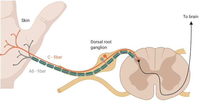
Aδ fibres are mechano-nociceptors, meaning that they are activated by high mechanical pressures. C fibres are polymodal, which means that they respond to a variety of noxious stimulations, such as noxious chemicals (e.g., acids), extreme temperatures and high mechanical pressures. They not only encode the stimulus modality (type), but also their intensity and duration, which are relayed to reflex centres in the central nervous sytem, mediating withdrawal reactions.

The nociceptive information travels from the DRG to different parts of the brain via spinothalamic tracts (from the spinal cord to the thalamus) and sensory fibres of the trigeminal tract (from the face to the thalamus). And it is within the brain that pain happens.

Pain is a complex feeling. Many brain areas are involved in not just generating pain, but also in ameliorating it. Structures from the limbic system, such as the amygdala, receive and integrate nociceptive and affect-related information. The amygdala can lead to increased nocifensive and affective pain behavior, while, under certain circumstances, it can also contribute to endogenous pain inhibition. Pain is also processed in the hypothalamus, the basal ganglia, the insula and the somatosensory cortices. Because these areas play a role in metabolism, as well as fear, pleasure and homeostasis, the nociceptive information is integrated and modulated according to the current state of the individual. In some situations, pain becomes pathological, as it is the case in neuropathic pain, where either previously innocuous stimuli become painful (aka, allodynia), or previously painful stimuli become even more painful (aka, hyperalgesia).

There are two brainstem structures, which are highly involved in controlling pain and generating analgesia. One of them is the periaqueductal grey (PAG) and the other is the rostral ventromedial medulla. These regions exert control over pain to prioritise competing stimuli, and to maintain homeostasis and survival. You might have noticed that, in highly stressful situations you do not feel pain. This is known as stress-induced analgesia, a phenomenon whereby the brain responds to stress by the production of endogenous opioids that act as natural analgesics in the nervous system. The opioid receptors found in the brain are the same ones which analgesic drugs, such as synthetic opioids and morphine, act on to relieve pain.

The evolution of pain

Many animal taxa have nociceptors. A schematic of the evolutionary development of nociceptors and the types of noxious stimuli they respond to is presented in Figure 3. In order to process nociceptive inputs, animals need a central nervous system (spinal cord and brain). It might come as a surprise that such a system, though at different levels of complexity, is found in all kinds of animals, including insects (like Drosophila melanogaster, the fruit fly), C. elegans (a type of worm highly studied in neurosciences), fish, amphibians, reptiles, birds and, of course, mammals.

Figure 3. The different types of nociceptors across animal taxa, from an evolutionary perspective.
Taken from Sneddon (2017)

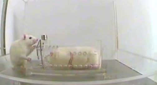
Life-history shapes pain perception. A very interesting example is the African naked mole rat, which lives in underground burrows that are poorly ventilated, hence contain high carbon dioxide levels. As a result, the C fibres of the naked mole rat are unresponsive to acid, which means that, while other mammals find acidic environments nociceptive, the African naked mole rat does not.

When it comes to acquatic animals, such as manatees, they are expected to have differences in their sensory system compared to terrestrial ones, due to distinct ecological and evolutionary pressures. In water, any chemicals become dilluted, shifts in temperatures are less common, and there is no mechanical damage due to falling. Thus, acquatic animals are possibly at a lower risk of damage than terrestrial animals, which has implications on their nociceptive system.

As far as manatees go, it is still unclear to what extent they feel pain. The fact that they are an endangered species makes is difficult to study them. But given that they posses a very well-developed somatosensory system, which is even more advanced than in other mammals, it is expected that manatees are familiar with pain. Moreover, we still do not know enough about their stress, fear, memory and pleasure systems, which all play a role in pain processing.

It would be great if we managed to achieve a better understanding of these amazing marine animals. But, until then, let us enjoy their existance peacefully, without interfering violently with their lifestyles and without exposing them to any potential pains.

For a more in-depth view on pain, as well as more information about manatees, I highly encourage you to read the papers and articles listed in References.

Special thanks to Isuru Priyaranga for creating the cover image. He is a fellow blogger and YouTuber, and I highly recommend visiting his blog and YouTube Channel.

References

Forced to suffer for science: From animal cruelty and experimental inefficiency to a change of perspective.

We, as scientists, have become desensitised to the pain, the distress and the physical and emotional damage that we inflict on laboratory animals. So much so, that we constantly find justifications for our cruel experiments in the goal of finding cures for the illnesses of our conspecifics, and in the rules and regulations that authorise these heartless procedures.

Despite ongoing widespread use of animal models in research, recently there has been extensive criticism on the state of drug development in psychiatry, calling for a switch from rodent behavioral pharmacology to mechanistic studies in cellular systems. In a recent paper, Heilig and colleagues argue that:

Overall, neuroscience has simply had very little impact on clinical alcoholism treatment. The situation is representative of a broader translational crisis in psychiatric neuroscience. Because translational failures in this area have been the rule rather than the exception, pharmaceutical industry has largely retracted from efforts to develop novel psychiatric medication. As a result, the utility of animal models in research on psychiatric disorders, including addiction, is also being questioned.

Heilig et al. (2019)

Caricature Cruelty

Several experimental paradigms employed by labs all over the world, for elucidating the mechanisms of mental disorders and for the development of new psychiatric drugs, consist of procedures that innevitably cause suffering to the experimental animals. From learned helplessness paradigms (forced swim and tail suspension), intended to model the symptoms of depression in humans, to neuropathic pain models, which involve nerve operations to induce chronic pain in rats or mice, as well as fear conditioning experiments, consisting of series of electric shocks on consecutive days, large numbers of laboratory animals across the globe are subjected to procedures at the end of which they are euthanized for histological analyses.

The two videos below illustrate two paradigms for learned helplessness in rodents – forced swim and tail suspension, respectively. Even for those unfamiliar with these methods, it is not hard to notice the amount of distress and fear the animals are forced to go through.

Another example, otherwise claimed to be minimally invasive and highly relevant for medication testing (Meinhardt and Sommer, 2015), is the post-dependent animal model, a model for medication development in alcoholism. It involves inducing dependence through repeated intermittent cycles of alcohol vapour exposure. In other words, rodents (usually, rats) are exposed every day, for several weeks or months, to cycles of intoxication with alcohol vapours, alternating with withdrawal, which ultimately result in compulsive alcohol intake, excessive alcohol seeking, hypersensitivity to stress as well as the development of an alcohol withdrawal syndrome, which better resemble human alcoholism.

Rats usually undergo 5 cycles of 14 (sometimes, 16) hours of forced exposure to alcohol vapours, separated by 10-hour periods of withdrawal and an additional 58 hours at the end of each weekly cycle. These cycles take place over many weeks. As a result of severe alcohol intoxication, some rats die during the experiment. At the end of the last alcohol exposure, the rats that have survived are decapitated.

The sardonically humorous caricatures below, selected from (Meinhardt and Sommer, 2015), not only illustrate the procedure, but are at the same time indicative of a certain emotional detachement these scientists have developed from the rats they used in their experiments.

Mainhardt & Sommer (2015)

“Unavoidable” Suffering

Granted, there have been attempts at reducing the suffering of these poor animals. The three Rs – Replacement, Reduction and Refinement – reflect the scope to encourage alternatives to animal testing, as well as improving animal welfare in experiments where the use of animals is unavoidable. The 3Rs have been incorporated into the legislation governing animal use in many countries, in order to ensure that the use of animals in testing is as ethical as possible.

And yet, with paradigms such as forced swimming test, also known as the behavioural despair test, or the tail suspension test ( where the rodent is hanging from its tail upside down and is unable to touch the walls of the compartment), it is clear that there is a big discrepancy between what could be done and what is actually being done. We could move away from these cruel practices, which have been demonstrated to be misleading and offer little understanding on the mechanisms behind psychiatric conditions, and, instead, resort to alternative strategies, which have the potential to set research on a path of true breakthroughs in psychiatry (as it is being presented in a later section). However, most papers focused on schizophrenia, anxiety, depression, Alzheimer’s disease, addiction etc. rely on experiments which would make the skin of the more sensitive of us crawl, and those with a tougher skin reconsider their academic career (such as switching to cognitive neuroscience and human-based studies only, in my case).

It is also important to remember that, not only the procedures themselves cause pain and suffering to the experimental animals, but often times the side-effects of the medications being tested on them and the behavioral tests they are being used in result in long-term health consequences – for instance, postdependent alcoholic rats end up developing peripheral secondary osteoporosis.

When suffering exceeds a certain limit, the animals are usually euthanized. What a great life these creatures must have, given that one of the best solutions to end their pain is premature death…


Rats empathise with other helpless rats

Although we all know that “animals have feelings too”, we are still far from understanding to what extent animals actually feel. In humans, for instance, pain and consciousness are tightly linked. We do not yet know which animals have consciousness and what (if anything) that consciousness might be like.

That being said, a study from 2011 demonstrated that rats exhibit emotional responses and empathy. In their experiment, Bartal and colleagues showed that when a free rat occasionally heard distress calls from another rat trapped in a cage, it learned to open the cage and released the other animal even in the absence of a payoff reunion with it. The free rat would even save a chocolate chip for the captive.


The presence of a rat trapped in a restrainer elicits focused activity from his cagemate, leading eventually to door-opening and consequent liberation of the trapped rat. (Science/AAAS).

This experiment clearly shows that rats, and possibly other animals as well, are capable of complex emotional experiences, previously only attributed to humans (more studies should be done to investigate this fascinating and important topic). Alas, in the absence of a deep understanding of the animal psyche, and moreover, with clear indications that animals possess the capacity to feel almost to the same extent as us humans do, we still continue to abuse them in our cruel experiments.


The issues with animal models

Valid disease models do not exist for psychiatric disorders.

Hyman (2012)

On the rodent models based on learned helplessness, Hyman went on to argue that:

Forced-swim and tail suspension tests do not even model the therapeutic action of antidepressants, because in those rodent screens a single dose of antidepressant is active, whereas in dependent patients, antidepressant drugs require weeks of administration to exert a therapeutic effect.

Hyman (2012)

In fact, given that most psychiatric diseases are heterogenous and polygenic, often times animal behavioral models have turned out to be misleading. Shockingly, only 8% of the CNS drug candidates developed between 1993 and 2004, which reached initial human testing, were approved to be used as medication. The main drawbacks of these drugs were the toxicity discovered in late-stage clinical trials, along with the inability to demonstrate efficacy. Not to mention the serious side effects these drugs produce in humans, such as weight gain and metabolic derrangements.

Animal models, albeit useful for some translational investigations and for basic studies in neuroscience, present various limitations:

  • Lack of molecular and neural circuit-based characteristics, which are required for molecular studies of psychiatric diseases.
  • The construction of transgenic mice is too slow and expensive.
  • Regarding non-human primates, the challenges involve cost, less well-developed technologies as well as ethical barriers.
  • When it comes to invertebrate models or zebrafish (extensively used in translational research), evolutionary distance poses huge obstacles in translational psychiatry, although they could be useful in the initial molecular investigations of the functions of risk alleles emerging from genetic studies.

Given the above-mentioned drawbacks of relying on animal models to develop psychiatric treatments, major pharmaceutical companies have already decided to move away from these old-fashioned approaches. Now, the question remains, what is there to do in the future?


Adopting new strategies in psychiatric research

Science needs to move forward and find better methods to study the highly complex mechanisms underlying psychiatric diseases, in order to allow for truly efficient drugs and therapies to be developed. As mentioned earlier, animal-based studies have more often than not failed to identify pharmaceutical compounds with positive outcomes in humans.

Over the last half-century, despite the identification of several antipsychotic and antidepressant drugs, alongside the discovery of various neurotransmitters, receptors and transporters involved in mental illnesses, objective diagnostic scans are scarce, and, surprisingly, only a handful of validated molecular targets have been established.

Luckily for us, there are several alternative solutions, which are already being seriously considered by various laboratories and drug companies, as listed below:

  • DNA sequencing, which is nowadays much cheaper than it used to be (by 1 million-fold), makes it possible to analyse large number of subjects, in the attempt to identify genes involved in the heterogenous, polygenic psychiatric disorders.
  • Large scale studies of gene function, epigenetics, transcriptomics and proteomics would contribute to the understanding of pathogenesis.
  • Optogenetics, a technology with increasing popularity in neurobiology, allows researchers to activate or inhibit single cell types, thus detecting which circuits are specific to certain disorders.
  • Human neurones derived directly from skin fibroblasts and blood cells in vitro, or generated from human embryonic stem cells (hESCs) or induced pluripotent stem cells (iPSCs) in vitro.
  • These above-mentioned tools can be combined with electrophysiological and neuroimaging data from humans, which can indirectly reveal abnormal functioning of widely distributed circuits.

What psychiatric research needs is to be able to accurately model molecular mechanisms of disease, instead of relying on behavioral results. Since I already mentioned large-scale genetic as well as epigenetic strategies, it is fair to admit that such studies require suitable living systems in which experiments can be conducted (given that the living human brain is not accessible, and that postmortem studies have limitations when it comes to functional analyses). Although, in some circumstances, animal behavioral experiments can help in elucidating treatment options, conclusions ought not to be based on modelling disease symptoms, as these can be misleading and often fail to translate into human psychopathology. Moreover, symptoms change over time and depending on the context, and are based on subjective rating scales, making the comparison between human and animal conditions difficult.

The solution is plain and simple – scientists and pharmaceutical companies must, first of all, unanimously and once and for all come to terms with the fact that the efforts based on cruel animal studies have been of too little avail to justify their continuation. Instead, a new strategy must be incorporated by the scientific community in psychiatric research, which should carry on from cell-based models and established molecular mechanisms to early human trials, skipping the intermediate step of animal behavioral models.

To end on a cheerful note, here is a heartwarming video which proves there is hope that the future could look bright for laboratory animals if people are willing to start making a change:


Special thanks to my mom for insightful comments and for her constant support, and to Gasser Elmissiery for inspiring discussions and for his contribution to creating the featured image.

References

  1. Bartal, I. B.-A., Decety, J., & Mason, P. (2011). Empathy and Pro-Social Behavior in Rats. Science334(6061), 1427 LP – 1430. doi:org/10.1126/science.1210789
  2. Haaranen M, Scuppa G, Tambalo S, Järvi V, Bertozzi SM, Armirotti A, Sommer WH, Bifone A, Hyytiä P. (2020). Anterior insula stimulation suppresses appetitive behavior while inducing forebrain activation in alcohol-preferring rats. Transl Psychiatry. 10(1):150. doi: 10.1038/s41398-020-0833-7
  3. Hansson AC, Koopmann A, Uhrig S, Bühler S, Domi E, Kiessling E, Ciccocioppo R, Froemke RC, Grinevich V, Kiefer F, Sommer WH, Vollstädt-Klein S, Spanagel R. (2018). Oxytocin Reduces Alcohol Cue-Reactivity in Alcohol-Dependent Rats and Humans. Neuropsychopharmacology. 43(6):1235-1246. doi: 10.1038/npp.2017.257
  4. Heilig M, Augier E, Pfarr S, Sommer WH. (2019). Developing neuroscience-based treatments for alcohol addiction: A matter of choice? Transl Psychiatry. 9(1):255. doi: 10.1038/s41398-019-0591-6
  5. Hyman SE. (2012). Revolution stalled. Sci Transl Med. 4:155cm11
  6. Knobloch HS, Charlet A, Hoffmann LC, Eliava M, Khrulev S, Cetin AH, Osten P, Schwarz MK, Seeburg PH, Stoop R, Grinevich V. (2012). Evoked axonal oxytocin release in the central amygdala attenuates fear response. Neuron. 73(3):553-66. doi: 10.1016/j.neuron.2011.11.030
  7. Meinhardt MW, Sommer WH. Postdependent state in rats as a model for medication development in alcoholism (2015). Addict Biol. 20(1):1-21. doi: 10.1111/adb.12187
  8. Wahis, J., Kerspern, D., Althammer, F., Baudon, A., Goyon, S., Hagiwara, D., … Charlet, A. (2020). Oxytocin Acts on Astrocytes in the Central Amygdala to Promote a Positive Emotional State. BioRxiv, 2020.02.25.963884. doi:org/10.1101/2020.02.25.963884